

Experimental study on deep ultraviolet-C LED disinfection device for often-touch surfaces of advanced medical equipment in hospitals
Baogen Huang¹, Feng Wang¹, Jing Li², Suping Sun¹, Huiying Lv¹, Lu Lu¹, Quan Shi¹, Chenchen Ding¹ and Yiping Mao²*
¹Fengxian People’s Hospital, Xuzhou, Jiangsu, China, ²The Affiliated Hospital of Xuzhou Medical University, Xuzhou, China

Main text
Figures
Tables
References
Abstract
Objective: To evaluate the disinfection efficacy of a third-generation gallium nitride (GaN)-based deep ultraviolet (UVC) LED disinfection cabinet on often-touch surfaces of small advanced electronic devices in hospital clinical environments, and to provide evidence for its application in hospital infection control.
Methods: A total of 329 device surfaces from 10 randomly selected departments were sampled: 66 computer mice, 151 mobile phones, 53 Personal Digital Assistants (PDAs), 29 electronic glucometers, and 30 electronic sphygmomanometers. Pre-disinfection bacterial contamination levels were assessed. The UVC-LED cabinet was used for disinfection (10-min cycle, 24 mJ/cm2 dose), and a natural bacteria eradication test was conducted.
Results: A total of 658 sampling points (329 pre- and post-disinfection) were monitored. Before disinfection, 57.1% of device surfaces showed microbial contamination, with mice and cleaning staff's mobile phones exhibiting relatively severe bacterial contamination. After disinfection, 97.9% of device surfaces showed bacterial colony counts reduced to 0 CFU, with an average natural bacteria eradication rate of 99.9% (range ≥90%).
Conclusion: High-frequency use of small advanced electronic devices by healthcare workers poses hospital infection risks. Deep LED Ultraviolet-C disinfection effectively reduces microbial load on surfaces of devices incompatible with chemical disinfectants or heat. It is recommended as a routine disinfection measure for hospital infection control. This study demonstrates high efficacy under controlled conditions; however, real-world effectiveness may be influenced by factors such as device positioning and surface shadows.
Keywords: High-flow water disinfection; UVC LED, UVC irradiation; 5-log inactivation; Escherichia coli; Ozone-free
2.1 Study design
A prospective, controlled before-and-after study design was employed to evaluate the disinfection efficacy of the UVC-LED cabinet on naturally contaminated device surfaces under field conditions in a tertiary hospital.
2.2 Experimental materials
LED ultraviolet disinfection cabinet, parameters: internal dimensions (length 430 mm × width 365 mm × height 630 mm), LED ultraviolet lamp bead model LY-DUV-TB (size: 3.5 × 3.5 mm), continuous working lifespan: 10,000+ hours, primary wavelength 270~280 nm, intensity 40 μW/cm2 (monitored every 6 months). The light distribution and disinfection point layout are illustrated in Figures 1A–C.

(A) LED ultraviolet light distribution curve. (B) LED ultraviolet disinfection cabinet device. (C) Disinfection point simulation image. (D) Sampling area example before and after disinfection. (E) Microbial culture and identification of equipment surfaces revealed the distribution of 13 bacterial species (n = 140). (F) Sampling results of surface pollution of different equipment before disinfection (%).
Sample collection, culture, and identification: 0.9% sterile saline sampling solution, Sterile cotton swabs, Tryptic Soy Agar (TSA) medium, Constant-temperature incubator, Automated microbial mass spectrometer (Model Autof ms1000), etc.
2.3 Study subjects and sampling strategy
Inclusion criteria: Inpatient departments with ≥20 beds in use or high-priority departments for infection control. Using stratified random sampling, 10 departments were selected: three internal medicine departments (Rehabilitation Medicine, Radiation Oncology, Gastroenterology); four surgical departments (Neurosurgery, Orthopedics, General Surgery, Thoracic Surgery); three key infection control departments ( Intensive Care Unit (ICU), Hemodialysis Center, Catheterization Laboratory).
Based on the pre-disinfection surface sampling assessment records (Appendix 1), a purposive sampling strategy was used to select representative, frequently-touched device surfaces from each department, including computer mice, PDAs, glucometers, smartphones, and electronic blood pressure monitors. This approach ensured the inclusion of high-risk surfaces relevant to clinical practice, though it may limit generalizability.
Sampling was performed on the right front, side, and back surfaces of devices before disinfection (after peak clinical activity) and on the left front, side, and back surfaces after disinfection (see Figure 1D for sampling zones). This method was employed to avoid potential cross-contamination between pre- and post-disinfection samples; however, it assumes uniform contamination distribution, which may introduce measurement bias if surfaces are not homogeneously contaminated.
A total of 658 device surfaces (329 pre-and post-disinfection) were sampled: 66 computer mice, 151 smartphones, 53 PDAs, 29 glucometers, 30 blood pressure monitors. The sample size was determined based on departmental availability and the goal of obtaining a representative profile of contamination across major device types and clinical areas, rather than a formal power calculation.
2.4 Experimental methods
Disinfection process evaluation: The cabinet's disinfection cycle was standardized at 10-min per device (UV dose 24 mJ/cm2), on-site observed and record the process (Appendix 2).
Disinfection efficacy evaluation: Appendix 3 (UV Disinfection Efficacy Evaluation and Testing Record Form) was filled out. Samples were processed within 2 h: The sampling solution was vortexed, and 1.0 mL was inoculated onto TSA plates using the pour plate method (2 plates/sample).The pour plate method was selected for its ability to capture both surface-adherent and embedded microorganisms, and its alignment with standard microbiological protocols for disinfection efficacy testing in hospital settings (10); negative control were included. Plates were incubated at 35~37°C for 48 hours, bacterial identification was performed using an automated microbial mass spectrometer.
Disinfection efficacy formula: Kill rate (%) = (A − B) / A × 100 , where A = Pre-disinfection colony count (CFU/sample), B = Post-disinfection colony count (CFU/sample). Criteria for qualified disinfection: Natural bacteria kill rate ≥90% (log reduction ≥1), with ≥90% of samples meeting this threshold; negative controls show no growth (10–12).
2.5 Statistical analysis
Data were organized in Excel2017 and analyzed using SPSS 23.0. Due to the highly skewed distribution of bacterial colony counts, data are presented as median and interquartile range (IQR) in addition to mean ± standard (x̄ ± s) deviation where appropriate. Non-parametric tests (Mann–Whitney U, Kruskal–Wallis) were used for comparisons of bacterial loads. Categorical data were described as counts and percentages (%). A P-value < 0.05 was considered statistically significant.
2.6 Ethical considerations
This study was approved by the Institutional Review Board (IRB) of [The Medical Ethics Committee of The Affiliated Hospital of Xuzhou Medical University] Approval No: [XYFY2025 - kL106 - 01]. Verbal informed consent was obtained from all healthcare workers whose personal devices (e.g., mobile phones) were sampled, with assurance of data anonymity and that the devices would not be harmed by the sampling or disinfection process.
3.1 Sampling and culture outcomes
TABLE 1 Sampling results and positive culture rates (%) by device type.
| Sampling time | Mouse | Cellphone | PDA | BP monitor | Glucometer | Total |
|---|---|---|---|---|---|---|
| Pre-disinfection | 62.1% (41/66) | 57.0% (86/151) | 66.0% (35/53) | 53.3% (16/30) | 34.5% (10/29) | 57.1% (188/329) |
| Post-disinfection | 7.3% (3/41) | 0% (0/86) | 0% (0/35) | 6.25% (1/16) | 0% (0/10) | 2.1% (4/188) |
Among 188 positive cultures, 13 species (140 strains) were identified. Dominant pathogens Staphylococci (52.9%): Staphylococcus hominis, S. epidermidis, S. haemolyticus, S. warneriBacillus spp.: Bacillus kochii, Paenibacillus cookii. Other isolates: Micrococcus spp., Escherichia coli, Pseudomonas spp., Klebsiella spp., Streptococcus spp., Corynebacterium spp., Acinetobacter spp. (Table 2 and Figure 1E).
| Microorganism | Strains (n=140) | Proportion (%) |
|---|---|---|
| G+ Staphylococcus hominis | 28 | 20.00 |
| G+ S. epidermidis | 19 | 13.57 |
| G+ S. haemolyticus | 17 | 12.14 |
| G- Bacillus kochii | 16 | 11.43 |
| G+ Micrococcus spp. | 13 | 9.29 |
| G- Escherichia coli | 11 | 7.86 |
| G+ S. warneri | 10 | 7.14 |
| G- Paenibacillus cookie | 8 | 5.71 |
| G- Pseudomonas spp. | 6 | 4.29 |
| G+ Streptococcus spp. | 4 | 2.86 |
| G- Klebsiella spp. | 3 | 2.14 |
| G- Acinetobacter spp. | 3 | 2.14 |
| G+ Corynebacterium spp. | 2 | 1.43 |
Computer mice showed the highest pre-disinfection colony counts (Median: 150 CFU/sample, IQR: 48–380; Mean ± SD: 338 ± 535 CFU/sample), with 12% of samples exceeding 500 CFU and 4.88% surpassing 1,000 CFU. The high standard deviation reflects the extreme skewness of the data, with a few heavily contaminated outliers. PDA devices, healthcare workers' smartphones, and glucometers showed no detectable bacterial growth post-disinfection (colony count = 0 CFU). Computer mice and electronic blood pressure monitors retained minimal residual bacteria post-disinfection (Median: 0 CFU/sample; Mean < 5 CFU/sample) (Table 3 and Figure 1F).
| Device type | Sample size | Pre-disinfection colony count (CFU/sample) | Post-disinfection colony count (CFU/sample) | Bacterial elimination rate (%) |
|---|---|---|---|---|
| Computer mice | 41 | 150 (48–380); 338 ± 535 | 0 (0–0); 1 ± 4 | 99.7 ± 1.2 |
| PDAs | 35 | 55 (30–95); 69 ± 50 | 0 (0–0); 0 | 100 |
| Healthcare workers' smartphones | 86 | 45 (20–95); 71 ± 84 | 0 (0–0); 0 | 100 |
| Glucometers | 10 | 55 (18–135); 103 ± 135 | 0 (0–0); 0 | 100 |
| Electronic BP monitors | 16 | 65 (35–105); 77 ± 57 | 0 (0–0); 1 ± 3 | 99.6 ± 1.5 |
| Department | Doctor sample size (n1 = 22) | Doctors' office | Nurse sample size (n2 = 19) | Nurses' station | t-value | P-value |
|---|---|---|---|---|---|---|
| Internal medicine | 8 | 619 ± 998 | 7 | 223 ± 164 | 1.11 | 0.30 |
| Rehabilitation medicine | − | 350 | 0 | − | − | − |
| Radiation oncology | 3 | 63 ± 42 | 3 | 220 ± 210 | 1.27 | 0.30 |
| Gastroenterology | 4 | 1,103 ± 1,295 | 4 | 225 ± 156 | 1.35 | 0.27 |
| Surgery | 11 | 373 ± 558 | 10 | 256 ± 160 | 0.67 | 0.51 |
| Neurosurgery | 3 | 827 ± 1,020 | 4 | 305 ± 184 | 0.88 | 0.46 |
| Orthopedics | 2 | 335 ± 163 | 1 | 100 | − | − |
| General surgery | 3 | 93 ± 78 | 3 | 160 ± 60 | 1.18 | 0.30 |
| Thoracic surgery | 3 | 223 ± 183 | 2 | 380 ± 170 | 0.98 | 0.42 |
| Key hospital infection departments | 3 | 143 ± 67 | 2 | 140 ± 57 | 0.05 | 0.96 |
| ICU | 1 | 160 | 0 | − | − | − |
| Hemodialysis center | 2 | 135 ± 92 | 0 | − | − | − |
| Catheterization room | 0 | − | 2 | 140 ± 57 | − | − |
| Position | Sample size (n) | Pre-disinfection colony count | Post-disinfection colony count | F-value | P-value |
|---|---|---|---|---|---|
| Doctors | 25 | 51 ± 47 | 0 | 15.2 | < 0.001 |
| Nurses | 25 | 51 ± 40 | 0 | ||
| Administrative staff | 16 | 26 ± 20 | 0 | ||
| Support/cleaning staff | 20 | 156 ± 126 | 0 |
4.1 Key findings and interpretation
This study confirmed significant microbial contamination (57.1% positivity) on often-touch electronic device surfaces in a hospital setting, aligning with international reports (4, 7). The predominance of Staphylococcus spp. and the presence of opportunistic pathogens like E. coli, Klebsiella spp., Pseudomonas spp., and Acinetobacter spp. underscore the potential role of these surfaces as reservoirs for nosocomial pathogen transmission (2, 13). The higher contamination on computer mice and support staff phones likely reflects differences in usage patterns, frequency of disinfection, and hand hygiene compliance, suggesting targeted interventions are needed for these high-touch fomites.
4.2 Efficacy of UVC-LED disinfection and practical implications
The UVC-LED disinfection cabinet demonstrated high efficacy, achieving a 100% eradication rate (0 CFU post-disinfection) on PDAs, healthcare worker smartphones, and glucometers, and an overall average eradication rate of 99.9%. These results are consistent with a growing body of evidence supporting the efficacy of UVC-LED technology against a broad spectrum of microorganisms (8, 9, 14–21). The minimal residual contamination (< 5 CFU/sample) found on computer mice and blood pressure monitor cuffs is likely attributable to shadowing effects in complex geometries (e.g., mouse wheels, folded cuff surfaces) (1, 9, 17, 22), highlighting a key operational consideration.
Compared to chemical disinfectants, which can be corrosive, require long contact times, and leave potentially harmful residues (5–8), and traditional mercury UV lamps, which contain hazardous materials and have operational limitations (5), the UVC-LED cabinet offers a dry, chemical-free, and user-friendly alternative. Its 10-min cycle time, instant start-up, and compatibility with delicate electronics present a significant practical advantage for integration into busy clinical workflows without damaging equipment (23–25).
4.3 Strengths, limitations, and research context
This study, as a component of the research project “Research and Development of Deep Ultraviolet LED Hospital Environment Disinfection Technology Based on Third-Generation Semiconductor Gallium Nitride and Multicenter Study on Disinfection Efficacy,” has the following strengths: First, advanced technology: it pioneers the application of GaN-based deep UVC-LED technology for disinfecting hospital equipment surfaces, validating its feasibility. Second, clinically relevant design: it assesses devices across multiple departments via field study and identifies natural microbiota profiles. Third, significant efficacy: an average eradication rate of 99.9% establishes a foundation for subsequent research. The limitations of this study simultaneously outline directions for further development within the project: First, the single-center design highlights the necessity of the subsequent “multicenter study” to verify generalizability. Second, the lack of long-term compatibility assessment is a crucial step for the technology's transition to mature application. Third, the direct impact on hospital infection rates remains unverified, requiring more rigorous studies to build a complete evidence chain. Thus, this study accomplishes a key step from technology development to single-center efficacy verification. Its limitations precisely provide a clear starting point and rationale for the project's subsequent phases: multicenter validation, long-term safety assessment, and benefit analysis.
The widespread integration of high-tech medical devices, while essential for modern healthcare, introduces significant nosocomial infection risks through contamination of often-touch surfaces. this study provides strong evidence supporting the effectiveness of UVC-LED technology in reducing microbial bioburden on these critical surfaces. Specifically, it confirms that a deep UVC-LED disinfection cabinet is a highly effective, user-friendly, and sustainable solution for mitigating microbial contamination across a variety of electronic device surfaces in hospitals. Its implementation can significantly enhance infection control protocols, especially where chemical disinfection or traditional UV methods are inadequate. Therefore, the adoption of this technology in clinical settings is recommended.
Data availability statement
The original contributions presented in the study are included in the article/Supplementary material, further inquiries can be directed to the corresponding author.
Author contributions
BH: Writing – original draft, Writing – review & editing, Data curation, Methodology, Conceptualization, Formal analysis, Project administration, Validation, Investigation, Funding acquisition, Resources, Visualization, Software. FW: Writing – review & editing, Methodology, Project administration, Validation, Investigation. JL: Writing – review & editing, Methodology, Project administration, Validation, Investigation. SS: Writing – review & editing, Methodology, Project administration, Validation, Investigation. HL: Writing – review & editing, Data curation, Methodology, Formal analysis, Validation, Investigation. LL: Writing – review & editing, Data curation, Methodology, Formal analysis, Validation, Investigation. QS: Writing – review & editing, Data curation, Methodology, Formal analysis, Validation, Investigation. CD: Writing – review & editing, Data curation, Methodology, Formal analysis, Validation, Investigation. YM: Writing – review & editing, Data curation, Methodology, Supervision, Conceptualization, Formal analysis, Project administration, Validation, Investigation, Funding acquisition, Resources.
Funding
The author(s) declared that financial support was received for this work and/or its publication. This research was supported by the Medical Research Project of Jiangsu Provincial Health Commission: Research and Development of Deep Ultraviolet LED Hospital Environment Disinfection Technology Based on Third-Generation Semiconductor Gallium Nitride and Multicenter Study on Disinfection Efficacy. Referenced in the “Notice on Announcing the Approved Medical Research Projects and Award-Winning New Technology Introduction Assessment Projects for the 2024 Fiscal Year by the Provincial Health Commission” (Su Wei Ke Jiao [2025] No. 1), Project No. M2024066.
Acknowledgments
We are grateful to the two hospitals for their support of this research. We also thank Xuzhou Liyu Advanced Technology Co., Ltd. for generously providing the ultraviolet disinfection cabinet used in this study.
Conflict of interest
The author(s) declared that this work was conducted in the absence of any commercial or financial relationships that could be construed as a potential conflict of interest.
Generative AI statement
The author(s) declared that generative AI was not used in the creation of this manuscript.
Any alternative text (alt text) provided alongside figures in this article has been generated by Frontiers with the support of artificial intelligence and reasonable efforts have been made to ensure accuracy, including review by the authors wherever possible. If you identify any issues, please contact us.
Publisher’s note
All claims expressed in this article are solely those of the authors and do not necessarily represent those of their affiliated organizations, or those of the publisher, the editors and the reviewers. Any product that may be evaluated in this article, or claim that may be made by its manufacturer, is not guaranteed or endorsed by the publisher.
1.Cremers-PijpersSvan RossumCDautzenbergMWertheimHTostmannAHopmanJ. Disinfecting hand-held electronic devices with UV-C in a healthcare setting. Infect Prev Pract. (2021) 3:10033. doi: 10.1016/j.infpip.2021.100133
2.BrowneKWhiteNMRussoPLChengACStewardsonAJMattersonGet al. Investigating the effect of enhanced cleaning and disinfection of shared medical equipment on health-care-associated infections in Australia (CLEEN): a stepped-wedge, cluster randomised, controlled trial. Lancet Infect Dis. (2024) 24:1347–56. doi: 10.1016/S1473-3099(24)00399-2
3.ZhuRYZhangYCTianL. Application progress of new ultraviolet disinfection technology. Shanghai J Prev Med. (2024) 36:823–9. (Chinese). doi: 10.19428/j.cnki.sjpm.2024.24519
4.LiRRWangYYZhangBR. Investigation on microbial contamination of mobile phones of medical staff in a tertiary hospital and observation on the efficacy of two disinfection methods. Chin J Disinfect. (2024) 41:954–6. (Chinese). doi: 10.11726/j.issn.1001-7658.2024.12.022
5.WenSSZuoWCZhouYYeQNQinDHCehnXD. Research status and development trend of ultraviolet disinfection technology. Opt Techn. (2020) 46: 664–70. (Chinese). doi: 10.13741/j.cnki.11-1879/o4.2020.06.006
6.ZhangGLiuHDWuHShengDCMaYTFanCYet al. Application of ultraviolet disinfection technology in water plants and engineering examples. Water Purif Technol. (2024) 43:196–203. (Chinese). doi: 10.15890/j.cnki.jsjs.2024.06.023
7.HesslingMHaagRSicksB. Review of microbial touchscreen contamination for the determination of reasonable ultraviolet disinfection doses. GMS Hyg Infect Control. (2021) 16:Doc30. doi: 10.3205/dgkh000401
8.NyangaresiPOQinYChenGZhangBLuYShenL. Effects of single and combined UV-LEDs on inactivation and subsequent reactivation of E. coli in water disinfection. Water Res. (2018) 147:331–41. doi: 10.1016/j.watres.2018.10.014
9.ZhangWLiXNLinJYMaCCShenJDuan H. Preliminary study on the disinfection effect of an LED ultraviolet disinfection cabinet. Chin J Disinfect. (2024) 41:881–4. (Chinese). doi: 10.11726/j.issn.1001-7658.2024.12.001
10.General Administration of Quality Supervision Inspection and Quarantine of the People's Republic of China. Hygienic Standard for Disinfection in Hospitals: GB15982-2012. Beijing: Standards Press of China (2012). (Chinese).
11.National Health Commission of the People's Republic of China. Hygienic Requirements for Ultraviolet Disinfectors: GB 28235-2020. Beijing: Standards Press of China (2020). (Chinese).
12.National Health Commission of the People's Republic of China. Standard for Evaluation of Field Disinfection During the COVID-19 Pandemic: WS/T 774-2021 (2021). (Chinese).
13.MitchellBGHallLWhiteNBarnettAGHaltonKPatersonDLet al. An environmental cleaning bundle and health-care-associated infections in hospitals (REACH): a multicentre, randomised trial. Lancet Infect Dis. (2019) 19:410–8. doi: 10.1016/S1473-3099(18)30714-X
14.SunZCZhuJCZhuTTQuXRLiNZhaoYXet al. Evaluation of the disinfection effect of high-energy pulsed ultraviolet disinfection equipment in medical institution settings. Chin J Prev Med. (2024) 58:857–61. (Chinese). doi: 10.3760/cma.j.cn112150-20231220-00471
15.ShaoZXWeiQHRenZShaSSFeiCNHanJ. Study on the disinfection effect of LED UV disinfection device and its influencing factors. Chin J Disinf. (2024) 41:481–4. (Chinese). doi: 10.11726/j.issn.1001-7658.2024.07.001
16.XiaGYXLuXQChenYYXuQLiWT. Experimental study on the bactericidal effect of LED ultraviolet lamps. Chin J Disinfect. (2017) 34:1–2. (Chinese). doi: 10.11726/j.issn.1001-7658.2017.01.001
17.ZhuZCTaoFXiaoLJingYFYouZSYuXD. Experimental study on drinking water disinfection for individual soldiers using deep ultraviolet LED device. Chin J Disinfect. (2020) 37:652–5. (Chinese). doi: 10.11726/j.issn.1001-7658.2020.09.004
18.RattanakulSOgumaK. Inactivation kinetics and efficiencies of UV-LEDs against Pseudomonas aeruginosa, Legionella pneumophila, and surrogate microorganisms. Water Res. (2018) 130:31–7. doi: 10.1016/j.watres.2017.11.047
19.WooHBeckSEBoczekLACarlsonKBrinkmanNELindenKGet al. Efficacy of inactivation of human enteroviruses by dual-wavelength germicidal ultraviolet (UV-C) light emitting diodes (LEDs). Water (Basel). (2019) 11:1131. doi: 10.3390/w11061131
20.LiuXShangXCaiQHuJ. Lab- and pilot- scale evaluation of bacterial inactivation and reactivation influenced by UV-LEDs arrangements in continuous flow water disinfection reactors. J Water Process Eng. (2023) 55:104093. doi: 10.1016/j.jwpe.2023.104093
21.LvLYLiZHLiangXHLuHYDuDTZhangGH. Observation on the disinfection effect of LED UV device on drinking water in field canteens. Chin J Disinfect. (2017) 34:72–4. (Chinese). doi: 10.11726/j.issn.1001-7658.2017.01.024
22.LiNNZhangBDengBQ. Study on the tailing effect of ultraviolet disinfection. Guangzhou Chem Industry. (2016) 44:58–60. (Chinese).
23.MalhotraSWlodarczykJKuoCNgoCGlucoftMSumulongIet al. Shining a light on the pathogenicity of health care providers' mobile phones: use of a novel ultraviolet-C wave disinfection device. Am J Infect Control. (2020) 48:1370–4. doi: 10.1016/j.ajic.2020.05.040
24.KeeneRChatterjeePJinadathaCWilliamsM. Using a handheld UV device for disinfection in the patient care environment: a descriptive qualitative study. Nurs Health Sci. (2023) 25:556–62. doi: 10.1111/nhs.13047
25.WuHMYangJKZhangYXWangYCDaiQChenY. Development of a positive pressure bio-protective hood disinfection cabinet and observation of its disinfection effect. Chin J Disinfect. (2024) 41:655–8. (Chinese). doi: 10.11726/j.issn.1001-7658.2024.09.006


Get in Touch
Recent Posts
-
 There’s nothing more unsettling than walking into your bedroom and being hit by a persistent musty odor. No matter how often you clean, wash the bedsheets, or open the windows, that damp, earthy smell lingers on. It can make even the most beautifully decorated room feel uncomfortable and unhygienic.
 -
 <!-- Meta Tags --> <meta title="UV Water Treatment for Wineries: Chemical-Free Disinfection Guide | Massphoton"> <meta name="description" content="Complete guide to UV water treatment for wineries. Learn how UV-C disinfection protects wine quality by eliminating bacteria, yeasts, and protozoa withou
 -
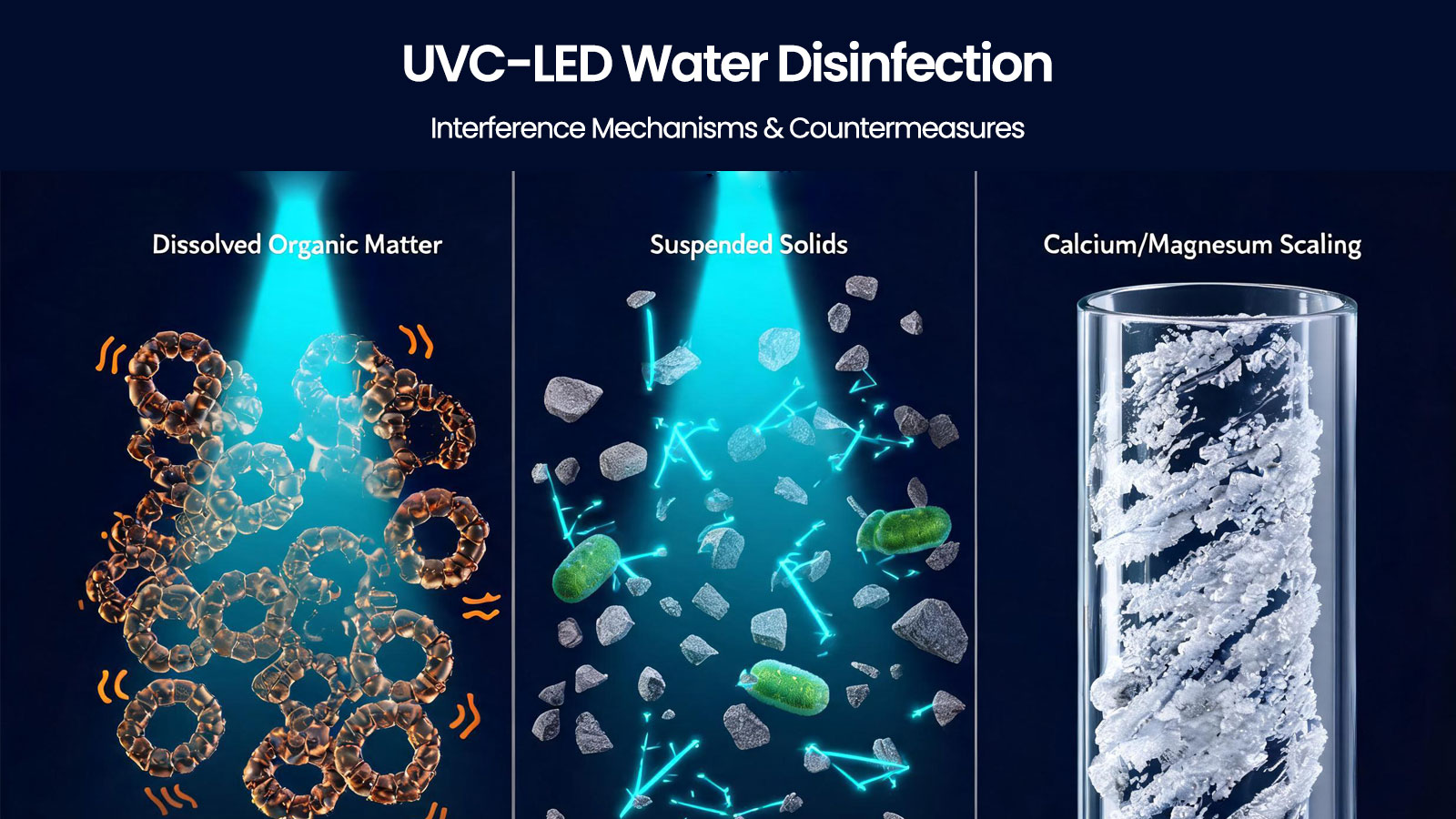 The content covers:1. Application advantages of UVC-LED for water disinfection2. Main interference factors in water and typical effects (with a data table)3. Prevention and control measures against water interference factors4. ConclusionI. Application Advantages of UVC-LED Water DisinfectionUVC LED
 -
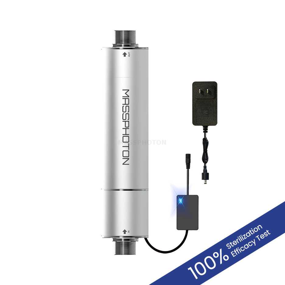 Deploying solid-state UVC LED water disinfection systems requires an accurate assessment of reactor hydrodynamics, targeted wavelengths, and source water quality variables like UV Transmittance (UVT). Unlike fragile, continuous-burn mercury lamps, deep UV LEDs offer space-saving footprints and instant on/off cycling that match real-world fluid dynamics. By coupling constant-current drivers with robust thermal management arrays, facility engineers can deliver precise germicidal doses to neutralize chlorine-resistant pathogens. This technical overview provides the sizing criteria, implementation strategies, and regulatory compliance paths needed to secure sustainable, chemical-free biosecurity.
